
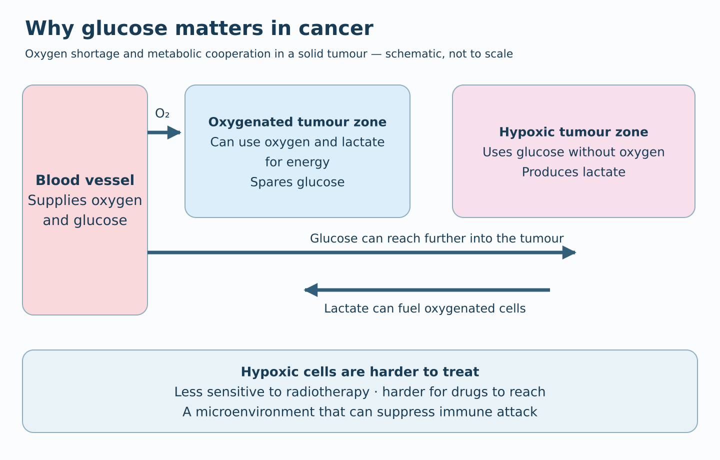
The figure shows how oxygen is rapidly consumed near blood vessels while glucose can reach further into a tumour. Oxygen-poor cells generate energy from glucose and produce lactate, which can then fuel better-oxygenated tumour cells.
Healthy cells and tumour cells live under different conditions
Healthy tissues have a well-developed network of small blood vessels supplying oxygen and nutrients.
With oxygen, cells can extract substantial energy from glucose, fat and other fuels.
Conditions in solid tumours are very different.
Tumour blood vessels are irregular, sparsely distributed and poorly functioning. Blood flow is uneven and may stop intermittently. Distances between vessels and tumour cells are also much greater than in healthy tissue.
As a result, oxygen delivery cannot meet the needs of all tumour cells.
This is called hypoxia — oxygen shortage — and is one of the most characteristic features of solid tumours.
These conditions are described by Vaupel and Harrison (2004).
Read Vaupel and Harrison on tumour hypoxia (2004) ↗
Oxygen runs out — but glucose can reach further
Oxygen and glucose arrive through the blood, but their availability within the tissue differs.
Cells near blood vessels continuously consume oxygen, so its concentration falls rapidly with distance.
Glucose is present at much higher concentrations and can reach further into tumour tissue.
This creates two regions with different conditions for energy production and survival.
Near blood vessels: tumour cells have oxygen and can use several fuels to produce energy.
Further from blood vessels: oxygen is limited, and cells increasingly depend on glucose for energy.
Differences in oxygen availability across tumour regions and their metabolic significance are discussed by Vaupel and Mayer (2017).
Read Vaupel and Mayer on tumour oxygenation (2017) ↗
Glucose has a special property: it can generate energy even without oxygen.
This process, glycolysis, yields far less energy per glucose molecule than oxidation with oxygen. However, glycolysis can operate rapidly and allow tumour cells to survive under conditions that would be very challenging for healthy cells.
For tumour cells experiencing severe oxygen shortage, glucose is therefore more than just one fuel among many. It is critical to survival.
Tumour cells cooperate over glucose and lactate
Perhaps the most fascinating aspect is that different tumour regions can cooperate like a connected organ to use limited resources.
When hypoxic tumour cells use glucose for energy, lactate is formed. Oxygen shortage limits their ability to use lactate as an oxidative fuel, so it is transported out of the cells.
Lactate was long regarded mainly as a metabolic waste product.
But lactate is also an excellent fuel, provided oxygen is available.
Tumour cells near blood vessels can therefore take up lactate from oxygen-poor regions and use it in their mitochondria to generate energy.
This creates cooperation:
- Oxygen-poor cells use glucose and produce lactate.
- Oxygenated cells use lactate as fuel, reducing their own glucose consumption.
- More glucose can then reach deeper into the tumour, where it is most needed.
Through the lactate it produces, glucose reaching hypoxic regions also contributes to energy supply elsewhere in the tumour.
A tumour does not need every cell to depend on glycolysis for glucose to be a central energy resource for the tissue as a whole.
This metabolic cooperation has been studied experimentally and was described particularly clearly by Sonveaux and colleagues (2008).
Read Sonveaux and colleagues on lactate-fuelled respiration (2008) ↗
An experiment revealing tumours' dependence on glucose
Sonveaux and colleagues investigated how oxygenated and oxygen-poor tumour cells use glucose and lactate.
They found that oxygenated tumour cells could use lactate as fuel and thereby spare glucose.
The researchers then blocked MCT1, a transport protein used by oxygenated tumour cells to take up lactate, disrupting cooperation between oxygenated and hypoxic regions.
The result was striking.
When oxygenated tumour cells could no longer use lactate, they consumed more glucose.
Less glucose consequently reached the hypoxic cells deeper in the tumour.
The oxygen-poor cells then died from glucose deprivation.
In animal experiments, tumour growth slowed, and the remaining tumour cells also became more sensitive to radiotherapy.
This elegantly illustrates how tumour metabolism can be targeted by disrupting cooperation between cell populations.
It also demonstrates the importance of glucose availability for survival in oxygen-poor tumour regions.
Read Sonveaux and colleagues: Targeting lactate-fueled respiration (2008) ↗
Cancer's paradox — the most treatment-resistant cells may be the most glucose-dependent
This brings us to something I find particularly interesting.
Hypoxic tumour regions contain some of the most difficult cancer cells to treat.
There are several explanations.
Radiotherapy: oxygen plays an important role in radiation's ability to produce lasting DNA damage. Radiation becomes less effective when oxygen is absent. Beckers, Pruschy and Vetrugno (2024) review hypoxia's contribution to radioresistance.
Read Beckers, Pruschy and Vetrugno on hypoxia and radiotherapy (2024) ↗
Chemotherapy: poor blood vessels and long transport distances make it difficult for drugs to reach oxygen-poor regions at sufficient concentrations. Hypoxic tumour cells also divide more slowly, reducing the effectiveness of drugs that primarily target rapidly dividing cells.
As early as 2002, Tannock and colleagues showed that several common chemotherapy drugs, including cisplatin, gemcitabine and paclitaxel, penetrated tumour-like tissue poorly. Cells far from vessels may therefore receive insufficient drug exposure even when they are intrinsically sensitive to the drug.
Read Tannock and colleagues on anticancer drug penetration (2002) ↗
Immunotherapy: oxygen shortage changes the tumour microenvironment. Lactate accumulation, altered pH and other metabolic adaptations impair immune cells' ability to attack the tumour.
Zhuang and colleagues (2023) discuss how hypoxia contributes to resistance to different treatments and the opportunities this creates for new treatment strategies.
Read Zhuang and colleagues on hypoxia signalling (2023) ↗
Oxygen-poor regions can thus act as refuges for cancer cells that survive treatment and later contribute to recurrence.
Yet these are precisely the regions where dependence on glucose is greatest.
The tumour cells that are hardest to treat with established therapies may also be the most dependent on glucose for survival.
This makes glucose metabolism a particularly interesting target for future cancer treatments.
Can we exploit tumours' glucose dependence?
If glucose is critical to energy supply in hypoxic tumour regions, a natural question follows:
What happens if we reduce glucose availability?
Several strategies are conceivable.
One is lowering blood glucose through dietary changes, such as the ketogenic diet discussed in the previous post.
Ketogenic diets and cancer — what does the research show?
Another is using drugs or other approaches to achieve a more pronounced, controlled reduction in blood glucose.
It is also possible to target cooperation between tumour zones, for example by blocking lactate transport, as in Sonveaux's experiments.
The central idea is the same: exploit the tumour's glucose dependence to target cells that are difficult to reach with traditional treatments.
The body nevertheless has several mechanisms for maintaining blood glucose. Even during a strict ketogenic diet, the liver continues producing glucose. A modest fall in blood glucose may therefore be insufficient to seriously disrupt tumour energy supply.
This is an important question for future research.
What can we conclude?
Tumour metabolism is much more complex than the idea that cancer cells simply prefer sugar.
Uneven oxygen delivery creates regions with very different conditions for energy production and survival.
Cells in oxygen-poor regions become strongly dependent on glucose, while better-oxygenated cells use other fuels, including lactate from hypoxic regions.
This cooperation helps glucose reach the cells that need it most.
At the same time, hypoxic tumour regions are particularly problematic for cancer treatment because radiotherapy, several forms of chemotherapy and immunotherapy work less well there.
The key point is not that all cancer cells use glucose in the same way. Rather, the tumour functions as a connected organ, in which hypoxia and metabolic cooperation make glucose a central resource for survival.
This is why I believe treatments affecting glucose availability and tumour metabolism deserve considerably more attention in cancer research.
What would you like to know more about?
Do you have questions about glucose, oxygen shortage, lactate or cancer? Please leave a comment.
Future posts can explore how much blood glucose needs to fall to affect tumour metabolism, and why the difference between ketogenic diets and more pronounced glucose lowering may matter.
Sture Hobro
References and further reading
1. Vaupel P, Harrison L (2004). Tumor hypoxia: causative factors, compensatory mechanisms, and cellular response. The Oncologist. Read the article
2. Sonveaux P et al. (2008). Targeting lactate-fueled respiration selectively kills hypoxic tumor cells in mice. Journal of Clinical Investigation. Read the article
3. Vaupel P, Mayer A (2017). Tumor Oxygenation Status: Facts and Fallacies. Advances in Experimental Medicine and Biology. Read the article
4. Zhuang Y et al. (2023). Hypoxia signaling in cancer: Implications for therapeutic interventions. MedComm. Read the article
5. Beckers C, Pruschy M, Vetrugno I (2024). Tumor hypoxia and radiotherapy: A major driver of resistance even for novel radiotherapy modalities. Seminars in Cancer Biology. Read the article
6. Tannock IF et al. (2002). Limited penetration of anticancer drugs through tumor tissue: a potential cause of resistance of solid tumors to chemotherapy. Clinical Cancer Research, 8, 878–884. Read the article
Comments are public. Please do not include sensitive health information about yourself or others. Comments are reviewed before publication.
Comments
How your comment is handled
Linbro Therapeutics AB is responsible for comments. Your display name, text and date are published if your comment is approved. Your ChatGPT identity is used for sign-in and spam prevention, but is not shown publicly. Pending comments are deleted within 90 days. Approved comments remain until removed or the comment service is discontinued. Do not include sensitive health information. Contact sture.hobro@linbrotherapeutics.com to request removal or ask about how comments are handled.