
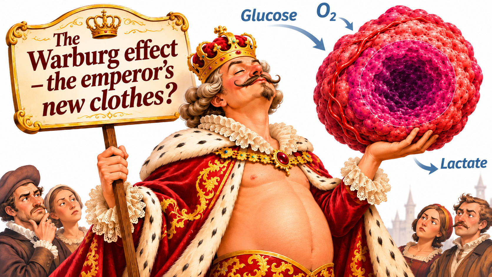
I am personally convinced that the classical Warburg effect plays no role in the energy metabolism of solid tumours.
That may seem a strange claim. For more than a hundred years, the Warburg effect has held an almost dogmatic position in cancer research. It is still described as a fundamental hallmark of cancer cells and is central to the account of cancer’s altered metabolism.
A Google Scholar search for “Warburg effect”, limited to publications since 2025, returned around 18,000 results. This is an estimate of search results rather than an exact count of scientific papers, but it illustrates the enormous attention the phenomenon continues to receive.
But does it really deserve this attention?
Behind this extensive scientific interest lies a fundamental question:
Where is the convincing evidence that aerobic glycolysis — the classical Warburg effect — is a dominant metabolic process in living solid tumours?
In Hans Christian Andersen’s fairy tale The Emperor’s New Clothes, everyone admired the emperor’s magnificent clothes. No one dared question what everyone else seemed to see. But when a child finally said what they saw, it became obvious that the emperor had no clothes.
I wonder whether there is a parallel in tumour metabolism.
There is extensive evidence that both cancer cells and healthy cells produce lactate despite ample oxygen in cell culture. The phenomenon is therefore not unique to cancer.
We also know that living tumours take up glucose and that some tumours are net exporters of lactate to the circulation.
But this is not the same as showing that well-oxygenated tumour cells mainly use glycolysis to produce ATP while converting large amounts of glucose to lactate.
What is missing is convincing evidence for the dominant role so often attributed to the Warburg effect in living tumours.
My view is that tumours’ glucose uptake and lactate production are explained by established biochemistry, limited oxygen availability and extensive metabolic cooperation between different parts of the tumour tissue.
What exactly is the Warburg effect?
In the 1920s, Otto Warburg observed that tumour tissue in laboratory experiments consumed glucose and formed large amounts of lactate even in the presence of oxygen.
This became known as aerobic glycolysis, or the Warburg effect.
To understand why this observation is remarkable, we need to distinguish between two ways of producing energy.
When oxygen is sufficiently available, cells use oxidative phosphorylation, OXPHOS, in their mitochondria. This makes very efficient use of the energy in glucose, fatty acids, lactate and other fuels.
When oxygen becomes insufficient, however, oxidative energy production is restricted. Cells then become more dependent on glycolysis, in which glucose is broken down to pyruvate while ATP is produced.
But for glycolysis to continue, NADH must be reoxidised to NAD⁺.
When oxygen shortage restricts mitochondrial respiration, the conversion of pyruvate to lactate by the enzyme lactate dehydrogenase, LDH, becomes crucial for regenerating NAD⁺.
This is hypoxic glycolysis — a normal and well-known adaptation to oxygen shortage.
The classical Warburg effect, by contrast, means that a cell produces substantial amounts of lactate despite having enough oxygen for oxidative energy production.
This distinction is crucial.
A tumour’s production of lactate does not prove that it exhibits the Warburg effect. The lactate may come from the tumour’s hypoxic regions.
Nor does a tumour’s uptake of large amounts of glucose tell us how much of its ATP is produced by glycolysis versus OXPHOS.
Zu and Guppy challenged the established view back in 2004
In 2004, Xin Lin Zu and Michael Guppy published Cancer metabolism: facts, fantasy, and fiction.
They reviewed forty years of research on cancer cell energy metabolism and questioned the idea that cancer cells generally have an intrinsic preference for glycolysis compared with healthy cells.
Their conclusion was clear: the research did not support the idea that cancer cells as a group are intrinsically glycolytic.
Instead, they argued that glycolysis in living tumours could be a consequence of their hypoxic environment.
This is a crucial distinction.
If a tumour cell uses glycolysis because oxygen is insufficient, we have not demonstrated the classical Warburg effect. We have demonstrated a normal metabolic adaptation to oxygen shortage.
More than twenty years ago, Zu and Guppy therefore identified a fundamental problem with the established account of cancer metabolism.
This also raises the question of how much of the aerobic glycolysis observed in the laboratory is actually a consequence of culture conditions.
Yet the traditional picture of the Warburg effect persists.
Healthy cells also exhibit the Warburg effect in culture
One particularly interesting observation is that extensive aerobic glycolysis is not unique to cultured cancer cells.
Healthy, proliferating cells also produce lactate despite the availability of oxygen.
This raises an obvious question:
If both healthy cells and cancer cells exhibit aerobic glycolysis in cell culture, how much of the phenomenon reflects cancer cell characteristics, and how much reflects the environment in which we grow them?
Cell cultures often use glucose concentrations of 5.5 or 25 mmol/L.
The lower concentration roughly corresponds to normal blood glucose, while the higher is about five times as high. Both exceed the low glucose concentrations measured in oxygen-poor parts of solid tumours.
The gas environment also normally contains considerably more oxygen than is found in large parts of living tumours. Actual oxygen availability at the cells is also affected by their density and oxygen consumption.
The culture medium is buffered to maintain a pH that favours cell growth, while lactate concentrations in fresh medium are considerably lower than in lactate-rich tumour regions.
Abundant glucose, good oxygen availability, relatively high pH and low lactate concentrations create favourable conditions for extensive glycolysis and lactate export.
These conditions differ from the oxygen-limited, lactate-rich and acidic environment in living tumours.
An abundant nutrient supply stimulates cellular energy metabolism and biosynthesis. At the same time, OXPHOS has a limited capacity, even when oxygen is available.
If glycolysis supplies pyruvate and NADH faster than oxidative metabolism can handle them, lactate production continues despite active mitochondria.
But high glucose uptake does not mean that OXPHOS is switched off.
As early as 2002, Guppy and colleagues showed that about 80% of ATP production in proliferating MCF-7 breast cancer cells came from oxidative processes and only 20% from glycolysis, under their experimental conditions.
That is a telling result.
Even cancer cells growing under favourable laboratory conditions thus produced most of their ATP through OXPHOS in this experiment.
But the researchers made another important observation.
The contribution of aerobic glycolysis in the MCF-7 cells did not differ from that in several non-transformed cell types examined.
What is usually described as a characteristic metabolic feature of cancer cells was therefore not even cancer-specific under these culture conditions.
Why, then, do such cells produce lactate despite available oxygen?
One explanation is that glycolysis supplies NADH faster than the cell’s other systems can reoxidise it. The capacity of oxidative metabolism also limits how much pyruvate can be used for further oxidation.
LDH then regenerates NAD⁺ by reducing pyruvate to lactate.
This is biochemically possible and has been demonstrated in cell models.
But the emergence of this metabolism in a culture dish is not proof that it plays a significant role in living tumours.
It is precisely this extrapolation from cell culture to living tumour tissue that I question.
A living tumour is not a culture dish
In solid tumours, the environment is radically different.
Blood vessels are unevenly distributed and often function poorly. Blood flow varies greatly between regions and over time.
Meanwhile, cells continuously consume oxygen and nutrients.
As a result, oxygen, glucose, lactate and other metabolites are distributed unevenly throughout the tissue.
Vaupel and his colleagues have documented these conditions over several decades, including in human tumours.
A central observation is that tumour cells retain substantial oxidative capacity, but oxygen availability restricts how much of that capacity is used.
In 2012, Vaupel and Mayer summarised this in an article with the telling title:
Availability, not respiratory capacity governs oxygen consumption of solid tumors.
In other words, in solid tumours it is largely oxygen availability, rather than the absence of functioning mitochondria, that limits respiration.
Another important feature is that, under typical tumour conditions, glucose reaches further from blood vessels than oxygen does.
This creates metabolic zonation.
Near functioning blood vessels, oxygen is available for OXPHOS.
Further from the vessels, oxygen shortage restricts oxidative energy production. Cells there become increasingly dependent on glycolysis for survival.
But these regions are not isolated from one another.
They are connected by the transport of glucose, lactate and other energy substrates.
This fundamentally changes how we need to understand tumour energy metabolism.
Sonveaux 2008 — demonstrating metabolic cooperation
In 2008, Pierre Sonveaux and colleagues published an experiment that I regard as one of the most important in modern tumour metabolism.
The researchers showed that oxygenated tumour cells use lactate as a fuel for oxidative phosphorylation.
This implied a completely different view of lactate’s function.
Rather than being merely a waste product of glycolysis, lactate serves as a valuable energy substrate.
The researchers described metabolic cooperation between the tumour’s oxygenated and hypoxic regions.
In hypoxic regions, glucose is used for glycolytic ATP production, with lactate formed at the same time.
The lactate is transported towards better-oxygenated regions, where tumour cells take it up through the transporter MCT1.
There, lactate is converted to pyruvate and used for oxidative energy production.
By using lactate, oxygenated cells reduce their glucose consumption.
More glucose can then reach the hypoxic cells that depend on glycolysis for their energy supply.
This means that different tumour regions do not merely compete for energy substrates. They also cooperate to use the limited supply of oxygen and glucose as efficiently as possible.
But Sonveaux did not stop at describing this cooperation.
He tested its importance experimentally.
What happened when lactate uptake was blocked?
The researchers blocked MCT1, preventing oxygenated tumour cells from using lactate in the same way.
Those cells then increased their glucose consumption and glycolysis.
Consequently, less glucose reached the hypoxic tumour regions.
Cells there suffered glucose deprivation and died.
By disrupting lactate uptake in the tumour’s oxygenated regions, the researchers could kill cells in its hypoxic regions.
This is a remarkable result.
The experiment showed that transport and metabolic cooperation have functional importance for tumour survival.
That same year, Gregg Semenza highlighted the findings in Tumor metabolism: cancer cells give and take lactate.
The title captures precisely what gets lost in the simplified account of the Warburg effect:
Tumour cells do not just produce lactate. They also use it as an oxidative fuel.
Read Sonveaux and colleagues (2008)
LDH — a reaction that cannot have net flux in both directions at once
Here lies another fundamental biochemical problem with the idea that the Warburg effect is the dominant metabolism in a tumour’s oxygenated regions.
The enzyme lactate dehydrogenase, LDH, catalyses the reaction:
Pyruvate + NADH + H⁺ ⇌ Lactate + NAD⁺
The reaction is reversible and close to equilibrium.
This means that its net direction is determined by the concentrations of lactate, pyruvate, NADH and NAD⁺, and by intracellular pH.
A high NADH/NAD⁺ ratio favours net lactate formation.
A high lactate concentration relative to pyruvate, by contrast, opposes further net lactate formation if other conditions remain unchanged.
There is a simple but important constraint here:
The same homogeneous intracellular LDH pool cannot simultaneously have a net flux from pyruvate to lactate and a net flux from lactate to pyruvate.
A car can move forwards or backwards, but hardly in both directions at once.
This does not mean that glycolysis and OXPHOS are mutually exclusive. A cell uses both processes simultaneously under many conditions.
Nor does it mean that LDH molecules only catalyse the reaction in one direction. Close to equilibrium, extensive exchange occurs in both directions.
But the net flux through a homogeneous reaction system has a definite direction.
If an oxygenated tumour cell is net oxidising lactate to pyruvate through LDH, that same LDH system cannot simultaneously have a net flux in the opposite direction.
It is therefore essential to distinguish glycolytic ATP production from the net lactate production of the classical Warburg phenotype.
Different intracellular environments or changes over time could theoretically permit more complex metabolic fluxes.
But such possibilities must be demonstrated before they are used to explain tumour metabolism.
To me, this makes Sonveaux’s findings more than an example of metabolic flexibility.
They pose a concrete problem for the assumption that oxygenated tumour cells are generally dominated by extensive net lactate production from glucose.
If lactate is consumed oxidatively in these cells, the same LDH system cannot simultaneously have the opposite net direction required for net lactate production in the classical Warburg phenotype.
High lactate and low pH — another constraint
The LDH reaction is only part of the problem.
For a cell to keep producing and exporting lactate, transport across its membrane must also function.
Lactate is transported mainly together with protons through monocarboxylate transporters, MCTs.
The net direction of transport is therefore determined by the concentration gradients of lactate and protons across the cell membrane.
In solid tumours, extracellular lactate is elevated and extracellular pH is often low.
These conditions oppose further lactate export. When the concentration gradients of lactate and protons no longer permit an outward net flux, export through the proton-coupled transporter ceases.
Tumour cells also normally maintain a higher intracellular than extracellular pH, which affects the direction of proton-coupled transport.
This means that extensive lactate production in a living tumour requires more than a high glycolytic flux.
Continued net formation and export also require a permissive redox driving force for LDH and suitable transport conditions.
Oxygen shortage becomes central here.
When insufficient oxygen restricts mitochondrial respiration, the mitochondria’s ability to contribute to the reoxidation of cytosolic NADH decreases.
The need to regenerate NAD⁺ through LDH then increases.
A higher NADH/NAD⁺ ratio drives the LDH reaction towards lactate formation.
Pyruvate is reduced to lactate, NAD⁺ is regenerated, and glycolysis continues producing ATP.
The central issue is therefore not an excess of pyruvate, but the cell’s need to regenerate NAD⁺ when mitochondrial oxidative metabolism is no longer sufficient.
This is why oxygen shortage is such a powerful driver of lactate production.
And since oxygen shortage is a fundamental feature of solid tumours, it provides a direct biochemical explanation for why tumours produce lactate.
That explanation does not require us to assume a general Warburg effect.
Sauer and Dauchy 1985 — a tumour changes its lactate flux within minutes
A study that I believe deserves considerably more attention was published in 1985 by Lawrence Sauer and Robert Dauchy.
The researchers studied living tumours in rats by measuring arteriovenous differences in substances including glucose and lactate.
This made it possible to determine whether the tumours were net consumers or releasers of these substances.
The results were fascinating.
In the tumour models examined, some tumours were net lactate producers while others were net consumers.
Among the Walker 256 tumours examined, 26% took up lactate from the blood. The corresponding proportion among Morris 5123C hepatomas was 48%.
Meanwhile, steady-state glucose consumption was similar in lactate-producing and lactate-consuming tumours.
But the most remarkable observation was that the very same tumour switched within minutes from net lactate production to net lactate uptake when the arterial blood lactate concentration was raised.
This demonstrates enormous metabolic flexibility.
Within a few minutes, a tumour can hardly rebuild its entire mitochondrial machinery or make extensive changes to its enzyme capacities.
Transport fluxes, substrate concentrations and reaction equilibria, however, change rapidly.
The findings therefore show how strongly a tumour’s metabolic behaviour is affected by its surroundings.
Sauer and Dauchy measured net flux across the whole tumour. The study therefore did not show that every individual cell changed its LDH direction.
The change could also have arisen from a shift in the balance between lactate-producing and lactate-consuming tumour regions.
But the main finding is equally interesting whichever mechanism dominated.
The very same living tumour switched within minutes between producing and consuming lactate when blood lactate changed. This shows how quickly tumour metabolism responds to altered substrate conditions.
It is difficult to reconcile this with the idea that extensive lactate production is a fixed, intrinsic property of cancer cells.
To me, Sauer and Dauchy’s findings are among the clearest examples of why tumour metabolism must be understood as a dynamic system.
Faubert and Hui 2017 — lactate as a fuel in living tumours too
Almost ten years after Sonveaux, two further important studies appeared.
Faubert and colleagues investigated metabolism in human lung tumours by using isotope-labelled energy substrates in patients.
They showed that carbon from circulating lactate was present in the tumours’ tricarboxylic acid, or TCA, cycle.
This was an important finding because it demonstrated that lactate contributes to oxidative metabolism in living human tumours too.
In complementary experimental tumour models in mice, the researchers went a step further. Direct comparisons between glucose and lactate showed that lactate contributed more carbon to the TCA cycle than glucose.
Lactate was therefore not merely an alternative fuel. In these tumour models, it was a more important carbon source for oxidative metabolism than glucose.
It was also particularly interesting that lactate use in the human lung tumours was evident in tumours with a high FDG-PET signal.
This means that a tumour with high uptake of the glucose analogue FDG also uses lactate as an oxidative substrate.
High FDG uptake is therefore not equivalent to a tumour mainly using glycolysis for its ATP production.
FDG is taken up by cells and phosphorylated by hexokinase. Unlike ordinary glucose, FDG-6-phosphate is not substantially metabolised further through glycolysis, but accumulates in the cells.
FDG-PET therefore does not directly measure glucose-to-lactate flux or the amount of ATP produced through glycolysis.
Faubert and colleagues also noted an important limitation.
Isotope labelling shows that lactate carbon reaches the TCA cycle, but does not automatically establish net lactate flux across the tumour.
The researchers could not perform a complete quantitative balance of how much glucose was taken up and how much lactate was exported from the human tumours.
This is particularly relevant to the question of the Warburg effect.
Without measuring net lactate production and relating it to local oxygen availability, it is impossible to determine how much of a tumour’s lactate production is genuinely aerobic.
Faubert also confirmed the importance of the lactate transporter MCT1 by genetically knocking it out in experimental mouse tumours. This greatly reduced lactate-dependent labelling of the tumours’ central metabolism.
That same year, Hui and colleagues showed that circulating lactate is a substantial carbon source for the TCA cycle in several tissues and experimental tumour models in mice.
This was another step away from the traditional view of lactate as merely a waste product.
Instead, lactate emerges as an important part of the exchange of energy substrates in the body and in tumours.
Together with Sonveaux, Semenza and Sauer, these studies provide a clear picture of dynamic tumour metabolism in which lactate is produced, transported and used as an oxidative substrate.
What does this mean for the Warburg effect?
Here we must return to the fundamental scientific question.
What has actually been demonstrated?
We know that both healthy cells and cancer cells produce lactate despite available oxygen in cell culture.
We know that tumours take up glucose.
We know that some tumours are net exporters of lactate.
We know that hypoxic cells become dependent on glycolytic ATP production when the oxygen supply cannot meet their energy needs.
We also know that tumour cells use lactate as an oxidative fuel, and that this forms part of functional metabolic cooperation between different tumour regions in experimentally studied tumours.
But where has it been convincingly demonstrated in a living human tumour that well-oxygenated cancer cells with preserved oxidative capacity convert large amounts of glucose to lactate on a net basis, and that this process constitutes a dominant part of the tumour’s energy metabolism?
A tumour’s production of lactate is not enough, because it may be due to hypoxia.
Its uptake of large amounts of glucose is not enough, because glucose is used in many other processes and is also oxidised.
Finding isotope-labelled glucose in lactate is not sufficient either to determine the local net flux or its energetic importance.
Nor does a cell exhibiting aerobic glycolysis in culture prove that the same process matters in a living tumour.
Moreover, oxidative lactate use has been demonstrated in oxygenated tumour cells.
As already noted, the same homogeneous LDH system cannot have two opposing net fluxes simultaneously.
Different cell populations can, of course, occupy different metabolic states. A cell can also change its metabolism over time.
But the existence of these possibilities is not itself proof that the Warburg effect plays a significant role in living tumours.
The burden of proof lies with those claiming that the classical Warburg effect is a general and dominant metabolic strategy in solid tumours.
To test this, researchers must distinguish aerobic from hypoxic lactate production, determine relevant net fluxes and estimate the share of tumour energy metabolism accounted for by these processes.
Only then can we establish whether the Warburg effect matters in living tumours.
Could the Warburg effect occur in some tumours after all?
Yes, it is biochemically possible, but it requires particular metabolic conditions.
Genetic or metabolic changes can restrict oxidative capacity in certain tumour types. If NADH reoxidation is then insufficient, LDH can contribute to continued net lactate production despite available oxygen.
Even well-oxygenated tumour cells could then produce measurable amounts of lactate.
However, it has not been proven that this is an important process in living human tumours, and my assessment is that in solid tumours it occurs at most marginally, if at all.
There is a fundamental difference between a mechanism being biochemically possible, being demonstrated in the laboratory and being a dominant metabolic process in living tumours.
Even if extensive aerobic glycolysis is demonstrated in a particular human tumour, it remains necessary to establish how much of the tumour is involved, how much ATP is produced this way and how widely the observation applies.
My assessment is that the classical Warburg effect, if it occurs in living tumours, is a very limited phenomenon associated with particular tumour types or metabolic conditions.
By contrast, there is an extensive experimental and biochemical basis for regarding hypoxic glycolysis, oxidative metabolism and the transport of energy substrates as central to solid tumours’ energy supply.
A tumour’s real metabolism rests on cooperation and competition
I believe one of the greatest weaknesses of the traditional account of the Warburg effect is its focus on the metabolic characteristics of individual cancer cells.
But a solid tumour is not merely a collection of isolated cells.
It resembles an organ more than we usually imagine.
It is a tissue in which different regions exist under entirely different conditions.
Oxygenated cells use OXPHOS for efficient ATP production.
Hypoxic cells become increasingly dependent on glycolysis.
Lactate is transported from producing to consuming cells, while glucose moves from blood vessels towards areas with the greatest need for glycolytic ATP production.
This allows different parts of the tumour to cooperate over limited energy resources.
Sauer showed how quickly a tumour’s net lactate flux changes when substrate availability changes.
Sonveaux showed how lactate transport and metabolic cooperation affect glucose supply and survival in hypoxic tumour regions.
Faubert and Hui showed that lactate contributes to oxidative metabolism, including in living tumours.
Meanwhile, Vaupel’s research shows how limited oxygen availability determines the conditions for tumour respiration.
Together, these findings describe a metabolism that is spatially organised, dependent on transport and highly flexible.
This does not mean that all tumours have exactly the same organisation. Tumour types differ in vascularisation, hypoxia, transport capacity and metabolic characteristics.
But we do not need to assume a universal preference for aerobic glycolysis to explain tumours’ glucose consumption and lactate production.
We already have an experimentally grounded alternative explanation.
Why does this matter for cancer treatment?
If the Warburg effect is regarded as cancer cells’ main energy metabolism, treatments risk being designed as though all tumour cells share the same metabolic dependence.
At the same time, the phenomenon is described as a preference for glycolysis — which is not the same as an absolute dependence.
If the tumour instead consists of metabolically distinct regions, both the treatment problem and the cells’ metabolic dependencies change.
Glucose has a special position because glycolysis provides ATP even without oxygen. In hypoxic regions, glucose and glycolysis are therefore crucial for cellular energy supply and survival. This is not a preference. It is a metabolic dependence.
An intervention affecting glucose availability has different consequences for hypoxic and oxygenated cells.
A treatment restricting lactate transport affects how energy substrates are distributed through the tumour tissue.
And a change in oxygen availability shifts the balance between glycolysis and OXPHOS without requiring a change in the cells’ underlying enzyme capacities.
Sonveaux’s experiment is an elegant example of how this can be exploited.
Preventing oxygenated tumour cells from using lactate increased their glucose consumption, depriving hypoxic cells of their energy substrate.
This is a very different approach to tumour metabolism from assuming that all cancer cells have an intrinsic preference for glycolysis.
It does not mean that inhibiting lactate or glucose transport is already an established cancer treatment. The path from an experimental mechanism to clinically effective treatment still needs to be tested.
But the central question is which metabolic processes the tumour actually needs to survive — not which processes cancer cells exhibit in a culture dish.
The emperor’s new clothes — what are we really seeing?
For more than a hundred years, the Warburg effect has held a special position in cancer research.
Today, we have extensive experimental evidence that tumours are metabolically heterogeneous, that their oxygen supply is uneven and that they use both glucose and lactate for energy.
We also have evidence that the transport of these substances connects different tumour regions and affects cell survival.
What I question is why the classical Warburg effect is still described as a dominant and almost self-evident feature of tumours when its quantitative importance in living human tumours has not been convincingly demonstrated.
I believe it is more reasonable to start from established biochemistry and documented conditions in tumour tissue.
Oxygen shortage restricts OXPHOS.
Glycolysis permits continued ATP production in hypoxic regions.
LDH regenerates NAD⁺ through lactate formation.
Lactate is transported and used as fuel in better-oxygenated areas.
This spares glucose for cells most dependent on glycolytic energy production.
This is more than a theoretical construction.
Important parts of this metabolic cooperation have been demonstrated experimentally, and the underlying biochemistry is well established.
My view is that this metabolic zonation, together with limited oxygen availability and energy substrate transport, describes a fundamental organising principle of solid tumours’ energy metabolism.
But the same standards of scientific evidence must also apply to the Warburg effect.
Today it is attributed a major role in tumour metabolism without its true quantitative importance in living human tumours having been established.
Anyone wishing to claim that aerobic glycolysis is a dominant energy process in living solid tumours must also demonstrate this — not just in a culture dish, where healthy cells exhibit the same phenomenon, but in the tumour tissue itself.
Perhaps it is time, like the child in Hans Christian Andersen’s fairy tale, to dare to ask the simple but uncomfortable question:
Where is the scientific evidence for the emperor’s clothes?
References
- Zu XL, Guppy M. (2004). Cancer metabolism: facts, fantasy, and fiction. Biochemical and Biophysical Research Communications, 313, 459–465. https://doi.org/10.1016/j.bbrc.2003.11.136
- Guppy M, Leedman P, Zu X, Russell V. (2002). Contribution by different fuels and metabolic pathways to the total ATP turnover of proliferating MCF-7 breast cancer cells. Biochemical Journal, 364, 309–315. https://doi.org/10.1042/bj3640309
- Sauer LA, Dauchy RT. (1985). Regulation of lactate production and utilization in rat tumors in vivo. Journal of Biological Chemistry, 260, 7496–7501. https://pubmed.ncbi.nlm.nih.gov/3997885/
- Sonveaux P, et al. (2008). Targeting lactate-fueled respiration selectively kills hypoxic tumor cells in mice. Journal of Clinical Investigation, 118, 3930–3942. https://doi.org/10.1172/JCI36843
- Semenza GL. (2008). Tumor metabolism: cancer cells give and take lactate. Journal of Clinical Investigation, 118, 3835–3837. https://doi.org/10.1172/JCI37373
- Vaupel P, Mayer A. (2012). Availability, not respiratory capacity governs oxygen consumption of solid tumors. International Journal of Biochemistry & Cell Biology, 44, 1477–1481. https://doi.org/10.1016/j.biocel.2012.05.019
- Faubert B, et al. (2017). Lactate metabolism in human lung tumors. Cell, 171, 358–371.e9. https://doi.org/10.1016/j.cell.2017.09.019
- Hui S, et al. (2017). Glucose feeds the TCA cycle via circulating lactate. Nature, 551, 115–118. https://doi.org/10.1038/nature24057
This article discusses scientific mechanisms and research findings. Its arguments are not treatment advice and do not imply that any established cancer treatment should be changed.
Comments are public. Please do not include sensitive health information about yourself or others. Comments are reviewed before publication.
Comments
How your comment is handled
Linbro Therapeutics AB is responsible for comments. Your display name, text and date are published if your comment is approved. Your ChatGPT identity is used for sign-in and spam prevention, but is not shown publicly. Pending comments are deleted within 90 days. Approved comments remain until removed or the comment service is discontinued. Do not include sensitive health information. Contact sture.hobro@linbrotherapeutics.com to request removal or ask about how comments are handled.